


Lipid Imaging Using Photoacoustic Remote Sensing Microscopy
Photoacoustic imaging, a state-of-the-art technology, utilizes light and sound to generate images of internal body structures. In this technique, a pulsed laser illuminates the biological tissue’s surface, leading to the absorption of photon energy, generating heat. The resulting thermoelastic expansion in the tissue releases energy in the form of ultrasonic waves. By scanning the sample and capturing corresponding photoacoustic signals, researchers can reconstruct 2D or 3D images of the biological tissue.
Typically, ultrasonic transducers are employed to collect these photoacoustic signals. However, due to the attenuation of sound waves in air, it is often necessary to introduce water or ultrasonic gel between the tissue sample and the transducer to ensure sensitive signal detection. This requirement for physical contact or immersion can significantly impact biological samples, restricting the practical application of conventional photoacoustic imaging in various scenarios.
On the contrary, ultrasonic transducers are constrained by their inherent structural and material properties, limiting their central response frequency and detection bandwidth. These limitations can diminish the sensitivity of traditional photoacoustic imaging systems when it comes to broadband signal detection. Recognizing these challenges, there is a need for an update to traditional photoacoustic imaging methods to enhance the quality of research in this field.
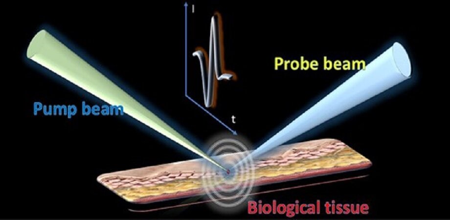
A promising advancement in photoacoustic imaging is the emergence of photoacoustic remote sensing. This novel modality deviates from the conventional approach of acoustic detection using ultrasonic transducers and instead employs another laser beam to detect acoustic signals. In this method, a separate laser source acts as the probe beam, confocal with the excitation beam.
When the sample absorbs energy, resulting in initial pressure, the instantaneous change in the sample surface’s refractive index, due to elasto-optical refractive index modulation, is monitored. The reflection intensity of the probe beam is observed to analyze the corresponding photoacoustic signals. This all-optical method of detecting acoustic signals eliminates the need for direct contact with the sample.
Additionally, optical sensing facilitates the transfer of detection bandwidth from the limited ultrasonic transducer to a broader photodiode, offering the potential for improved detection sensitivity and signal-to-noise ratio. Addressing these considerations, a research team from the University of Hong Kong recently introduced near-infrared photoacoustic remote sensing microscopy for non-contact imaging of lipids. Their approach employs a 1.7-μm thulium-doped fiber laser as the pump beam to selectively stimulate the C–H bond in lipids. Simultaneously, a 1.5-μm continuous-wave (CW) laser is utilized as the detection beam, confocal with the pump beam, to capture the initial ultrasonic pressure. This optical detection of ultrasonic signals eliminates the need for ultrasonic transducers and enables remote sensing of photoacoustic signals.
The method provides a broader detection bandwidth, enhancing detection sensitivity and signal-to-noise ratio. In their experimental demonstrations, the team showcased imaging results of two forms of pure lipid samples through photoacoustic remote sensing, analyzing the corresponding power spectrum density of the signals. The optical detection method demonstrated a broader frequency response compared to conventional transducers. The researchers also conducted photoacoustic remote sensing imaging on biological samples, such as nematodes and brain slices, revealing promising contrast and signal-to-noise ratios.
This showcases the high-performance imaging capabilities on the tissue scale. Corresponding author Kenneth K. Y. Wong, Professor of Engineering at the University of Hong Kong, notes, “Photoacoustic remote sensing microscopy achieves label-free imaging that can target specific molecular bonds. Optical detection of ultrasonic signals provides non-contact operation and a broader frequency response. Meanwhile, the photoacoustic remote sensing microscopy shows high performance for lipid distribution mapping on the tissue scale.” This innovative technique holds significant promise for diverse applications in biomedical research.
This article is republished from PhysORG under a Creative Commons license. Read the original article.
Do not forget to share your opinion with us to provide you with the best posts !


0 Comments